
Drs Decker and Damato following a T6 flight
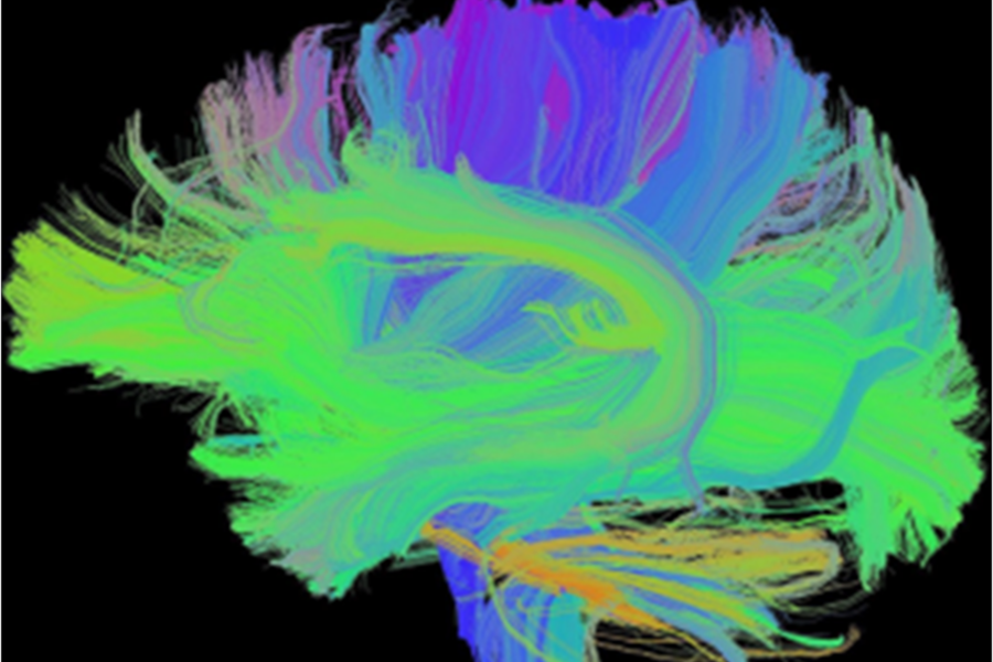
Digital analysis to visualize axonal tracts
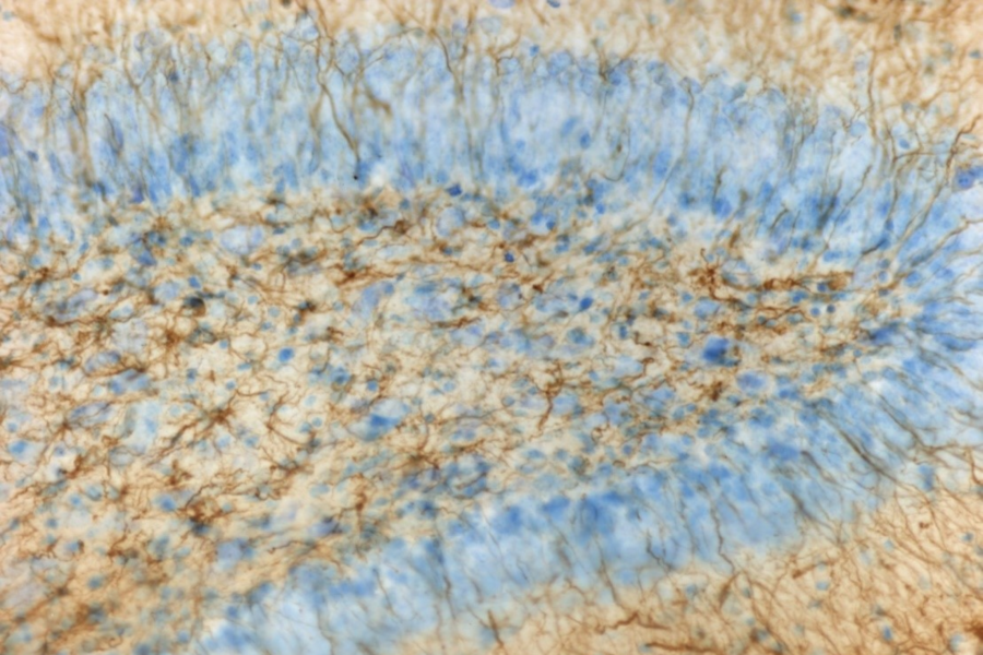
Immunohistochemistry analysis of rat hippocampus
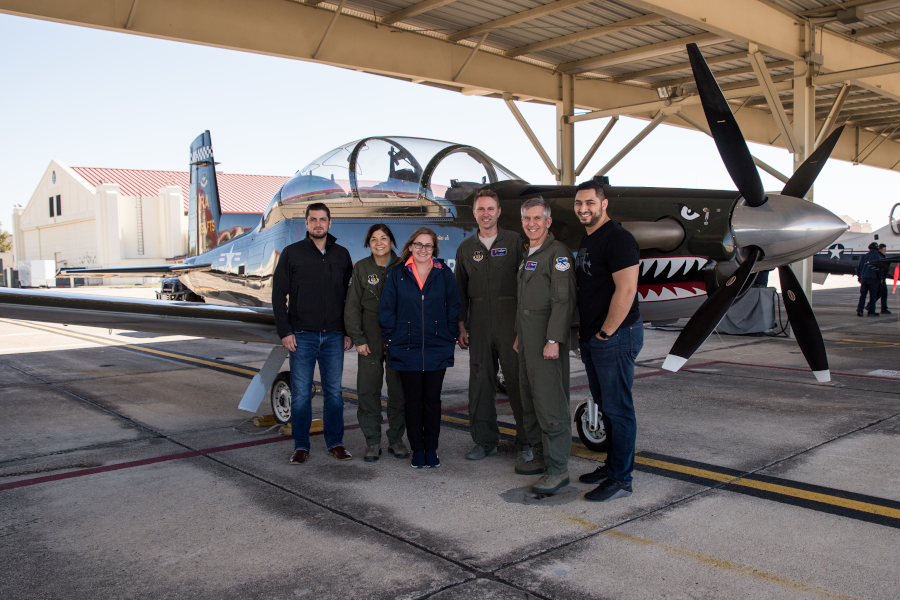
Our lab at Randolph AFB before Dr. Decker and Damato’s flight
Many factors jeopardize normal brain development. Disturbances to brain oxygenation and circulation are among the leading causes. The human brain is exquisitely sensitive to changes in the amount of oxygen that we breathe. Exposure to either low levels (hypoxia) or high levels (hyperoxia), if even for a brief duration, lead to immediate changes in brain function. Long term exposure to hypoxia or hyperoxia evokes changes in brain structure and function.
Clinical disorders can induce low oxygen levels in the bloodstream (hypoxemia). Examples include reduced cardiac output (cardiac disease, cardiogenic shock, etc.), lung disease, central nervous dysfunction (including drug overdose), premature birth accompanied by underdeveloped lungs, etc.
Environmental conditions can also lead to low levels of oxygen within the body. These include traveling through or living in high altitude environments (Leadville, Colorado; Tibetan Himalayan mountains; Bolivian Altiplano; the Ethiopian Simien Mountans). Certain occupations may also require that people live or work in slightly reduced oxygen environments to reduce fire hazards, such as submariners.
Throughout the course of evolution, humans have developed physiologic strategies to adapt to low oxygen levels. These include changes in ventilation and red blood cell mass. In sharp contrast, there is no place on Earth in which naturally occurring oxygen levels exceed 21%. Consequently, humans may not be able to adapt to high oxygen levels.
As high levels of oxygen are also a prerequisite to survival during high-speed, high altitude aviation, extravehicular activities around the international space station, or undersea diving, there is a growing need to understand the impact of hyperoxia upon brain structure and function.
Our ongoing program of research is focused upon characterizing structural and functional changes occurring within the human brain before, during and after exposure to hypo- and hyperoxia.
Funding Source: Department of Defense-Air Force Research Laboratories, FA8650-19-C-6103
Onboard oxygen generating systems (OBOGS) provide increased inspired oxygen (FiO2) to mitigate risk of hypobaric hypoxia and decompression injury to tactical aviators. However, FiO2 generated by OBOGS are not consistently delivered at fixed levels. Rather, FiO2 may be delivered oscillatory manner, with oxygen percentages ± 15% or more of prescribed values. The objective of this study was to characterize physiologic responses to exposure to both steady state and non-steady state inspired oxygen concentrations during normobaric and hypobaric environmental pressures. Primary outcomes include serial measurements of arterial blood gases and prefrontal cortical brain activity during cognitive challenge. Secondary outcomes include quantification of proinflammatory serum analytes within the circulatory system both before and after the exposures.
Funding Source: Department of Defense-US Navy; Naval Medical Research Unit-Dayton, N6264522C0010
Aviators are exposed to non-steady state levels of increased inspired oxygen (FiO2) through the aircraft’s onboard oxygen generating system. This study’s objective is to determine, in a controlled laboratory setting, if non-steady state inspired oxygen affects cortical electrical activity (measured through EEG) and cognitive performance in both normobaric and hypobaric conditions.
Defining mechanisms mediating vulnerability towards cognitive fatigue: Post Sortie Systemic Inflammation, Sleep, Diet, and Exercise
Funding Source: Department of Defense-Air Force Research Laboratories, 711th Human Performance Wing
A lack of evidence exists to confirm that oxygen delivery schedules, which determine FiO2 necessary to sustain appropriate PaO2 levels for tactical aviators, account for safety pressure, which could increase PaO2 over predicted values. The objective of this laboratory-based study is to measure PaO2 derived from specific FiO2 concentrations delivered with and without safety pressure. The overarching goal is to generate necessary specifications for positive pressure breathing systems designed to deliver FiO2 levels conferring maximal neuroprotection and resiliency within the cockpit, yet do not cross the critical threshold that triggers onset of potentially adverse outcomes.
Funding Source: Department of Defense-Air Force Research Laboratories, 711th Human Performance Wing
Cognitive fatigue, a constant threat to human performance and aviator safety, is the “likely cause of the next mishap” according to the National Commission on Military Aviation Safety, Report to the President and Congress. Preventing that mishap is hampered by an absence of quantitative physiologic biomarkers that predict increasing levels of cognitive fatigue. Our ongoing studies are addressing that critical knowledge gap. We have recently identified post sortie changes in serum levels of proinflammatory cytokines, proangiogenic proteins and neurotrophins in a cohort of T-6A Texan II Instructor Pilots (IPs). Those biochemical changes were also accompanied by reduced activity within the brain’s prefrontal cortex, an area critical for cognitive and executive processes including decision making and predicting consequences of current actions. Collectively, those novel findings are providing insight into intrinsic physiologic mechanisms corresponding with increasing levels of cognitive fatigue. This study’s objective is to define the specific causes initiating those biochemical and cognitive outcomes. Specifically, we will characterize the effects of increased concentrations of inspired oxygen and multiple hypergravity maneuvers on serum biochemical profiles, neuroinflammation, and patterns of cortical activity.
Disorientation, dizziness, and fatigue can rapidly occur, unprecipitated, during high-performance aviation. When unnoticed, the outcome can be catastrophic. Our ongoing studies are defining mechanisms that may contribute to the onset of those symptoms. The objective of this study was to characterize the neurovascular effects of breathing high concentrations of oxygen, a prerequisite during high-speed and very high altitude aviation, upon overall brain function. Our study found that high levels of inspired oxygen significantly reduced brain perfusion. This was accompanied by electroencephalographic (EEG) signatures of enhanced vigilance. We also found cognitive processes were heightened. Our data may suggest that the brain's "alerting" mechanisms are activated by a drop in perfusion. Results are published in:
Damato, E.G., Flak, T.A., Mayes, R.S., Strohl, K.P., Ziganti, A.M., Abdollahifar, A., Flask, C.A., LaManna, J.C., Decker, M.J. (2020). Neurovascular and cortical responses to hyperoxia: Enhanced cognition and electroencephalographic activity despite reduced perfusion. Journal of Physiology, 598 (18), 3941–3956. doi: 10.1113/JP279453
Funding Source: Air Force Research Laboratory (AFRL) under the Human Performance Aerospace Physiology Program (contract FA8650-17-F-6822, T.O. 0052).
Link for free text version: https://pubmed.ncbi.nlm.nih.gov/33174711/
Tactical aviation imposes unprecedented physical challenges including repetitive exposure to hypergravity, hyperoxia, increased work of breathing, and profound cognitive workloads. Each stressor evokes outcomes ranging from musculoskeletal duress and atelectasis to physical and cognitive fatigue, the latter among the foremost threats to aviators. Whereas sleep loss is traditionally considered the primary cause of fatigue in aviators, converging experimental, observational, and medical studies have identified biochemical mechanisms promoting onset of fatigue. Those mechanisms, which fundamentally differ from sleep loss, revolve around increased proinflammatory cytokines, produced and released in response to tissue injury, chronic inflammatory disorders, allergens, or physical duress. This study’s objective was to inform our understanding of potential relationships between serum levels of proinflammatory cytokines and onset of fatigue within a cohort of aviators who experience multiple high-performance sorties on a daily basis. While fatigue in aviators has been attributed almost solely to sleep loss, nocturnal sorties, or disrupted circadian rhythmicity, our study findings suggest an alternative mechanism that can promote onset of fatigue: increased blood levels of proinflammatory cytokines. Results are published in:
Damato, E.G., Fillioe, S.J., Mayes, R.S., Margevicius, S.P., Somogyi, J.E., Vannix, I.S., Abdollahifar, A., Turner, A.M., Ilcus, L.S., Decker, M.J. (2022). Increased serum levels of proinflammatory cytokines are accompanied by fatigue in military T-6A Texan II instructor pilots. Frontiers in Physiology, 13, 876750. doi: 10.3389/fphys.2022.876750
Funding Source: Department of Defense – 711th Human Performance Wing Studies and Analyses Program, F4F4FE0163G001
Link for free text version: https://pubmed.ncbi.nlm.nih.gov/35574470/
Tactical aviators require administration of enhanced inspired oxygen concentrations (hyperoxia) to reduce risk of hypobaric hypoxia and decompression injuries. Hyperoxia is not without consequence; it reduces cerebral perfusion (CBF). Characterizing the relationship between FiO2 and CBF is necessary to establish FiO2 levels that do not reduce CBF yet are sufficient to mitigate risk of in-flight physiological stressors. To achieve that goal, this study’s objective was to determine whether a dose-response relationship exists between FiO2 and CBF and, if so, the FiO2 at which CBF significantly declines. We found that the neurovascular system appears to respond to increasing FiO2 levels in a dose dependent manner, with significant reductions in CBF with FiO2 exposures ≥ 60%. Results are published in:
Damato, E.G., Fillioe, S.J., Vannix, I.S., Norton, L.K., Margevicius, S.P., Beebe, J.L., Decker, M.J. (2022). Characterizing the dose response of hyperoxia with brain perfusion. Aerospace Medicine & Human Performance, 93(6):493–498. doi: 10.3357/AMHP.6056.2022
Funding Source: Department of Defense-US Navy Bureau of Medicine and Surgery, N3239819P0321
Newborns exposed to hypoxia, especially in the days following birth, are at risk to become sleepy adolescents who are hyperactive while awake and also have poor working memory. By characterizing the neurochemical and neuroanatomic structure and function, we found suboptimal performance within the dopaminergic networks. Those studies formed the scientific structure premise for this project, employing magnetic resonance imaging, high-density electroencephalography, and other techniques to measure brain structure and function in children who were prematurely born and may have been exposed to hypoxia.
Funding Source: National Institutes of Health, R21NR017235-01